Differential language network functional connectivity alterations in Alzheimer's disease and the semantic variant of primary progressive aphasia

Résumé
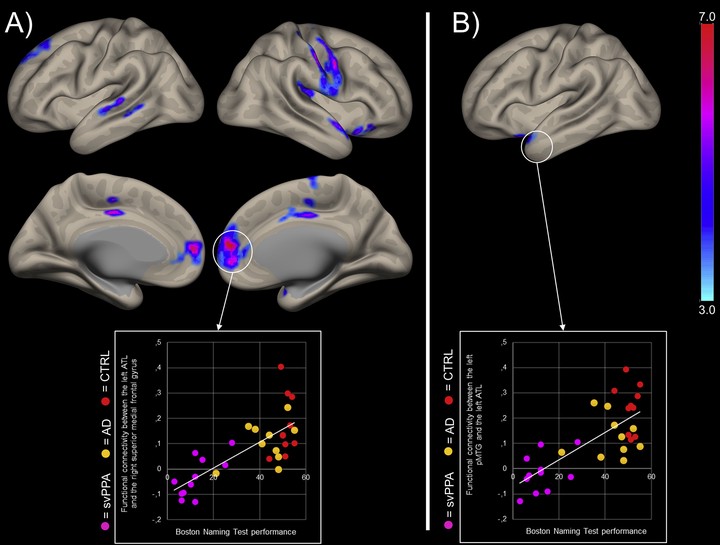
Patients with Alzheimer’s disease (AD) and semantic variant primary progressive aphasia (svPPA) can present with similar language impairments, mainly in naming. It has been hypothesized that these deficits are associated with different brain mechanisms in each disease, but no previous study has used a network approach to explore this hypothesis. The aim of this study was to compare resting-state functional magnetic resonance imaging (rs-fMRI) language network in AD, svPPA patients, and cognitively unimpaired elderly adults (CTRL). Therefore, 10 AD patients, 12 svPPA patients and 11 CTRL underwent rs-fMRI. Seed-based functional connectivity analyses were conducted using regions of interest in the left anterior temporal lobe (ATL), left posterior middle temporal gyrus (pMTG) and left inferior frontal gyrus (IFG), applying a voxelwise correction for gray matter volume. In AD patients, the left pMTG was the only key language region showing functional connectivity changes, mainly a reduced interhemispheric functional connectivity with its right-hemisphere counterpart, in comparison to CTRL. In svPPA patients, we observed a functional isolation of the left ATL, both decreases and increases in functional connectivity from the left pMTG and increased functional connectivity form the left IFG. Post-hoc analyses showed that naming impairments were overall associated with the functional disconnections observed across the language network. In conclusion, AD and svPPA patients present distinct language network functional connectivity profiles. In AD patients, functional connectivity changes were restricted to the left pMTG and were overall less severe in comparison to svPPA patients. Results in svPPA patients suggest decreased functional connectivity along the ventral language pathway and increased functional connectivity along the dorsal language pathway. Finally, the observed connectivity patterns are overall consistent with previously reported structural connectivity and language profiles in these patients.